
Sight and hearing in the context of the sensory stimulation and integration
Authors: MUDr. Martin Kučera, Mgr. Kateřina Fritzlová
Institut pro výzkum a léčbu poruch komunikace (Communication Disorder Research and Treatment Institute)
- Sight
1.1 Neuroanatomy and neurophysiology of visual perception
The sensory organ of sight is the eye. The visual system processes and deciphers large quantity of information, more than any afferent system of human body (Lowe, Webb). The basic perceived signal is light which is defined as electromagnetic wave. People visually perceive electromagnetic wave within the range of 390-790 nm (the range between ultra violet and infrared radiation). The basic characteristics of light are luminosity, frequency spectrum/colour, polarization and coherence. Light passes through the individual layers of the eyeball in the following order: cornea, anterior chamber, iris, posterior chamber, lens and vitreous humour. The basic function of the entire eyeball is to focus the image on the retina. Photoreceptors, rod and cone cells, which are also first neuron of the visual path, are located on the part of the retina averted from the globe. Cones provide for the colourful daytime (photopic) vision; rods provide for the night-time (scotopic) vision, which allows only the perception of changes in brightness (Čihák).
Image generated on the retina is inverted and rotated in the cerebral cortex. The visual path is partially crossed and consisting of four neurons. The first three neurons are located in the retina, axons of the third nerve form nervus opticus, after its crossing it is called tractus opticus. Alongside the tractus opticus there are branches providing reflex actions such as: mydriasa and miosa, lens accommodation and the convergence of globes. These functions are important for the image we perceive; they enable us to focus and merge images from right and left eye into one image. In addition, they permit for the maintenance of image during simultaneous movements of body and head (Synek, Skorková). The fourth neuron of the visual path ends in the primary cortex area of the occipital lobe (area 17, primary visual cortex). The signal is subsequently transmitted into the frontal eye field in the premotoric area of the frontal lobe; when processed an impulse is sent to move eyes. The impulse passes through the midbrain and the bridge into the reticular formation. The reticular formation sends impulses into the nuclei of extraocular muscles. Fibres coming from the medial (inner) side of the retina cross and the ones coming from the lateral (outer) side do not cross. This means that the image from the left half of each eye goes to the left hemisphere and the image from the right half of each eye goes to the right hemisphere.

1.1.1 Visual integration
From the point of view of visual perception processing, the visual association cortex in the area of temporal lobe is important. Damage to the association areas or the temporal lobe, or its connection with other parts of the brain, may have several impacts not only on the processing of visual perception, but also on the person’s movement and other functions, including the verbal function (Note: there are damages of: visual – auditory, visual – motoric, visual – somatic sensory and visual – verbal path). To determine and identify objects it is necessary to process the visualized object at all visual path levels (association area included) with the current integration of processed perceptions from other senses with the connection to further mental activity, including their integration with past experience (Mesulam, 1985 in Lowe, Webb).
Sight is an important sense which allows for spatial orientation of the body. There is an important relation between sight and memory and learning. If a person is exposed to various stimuli and uses sight at the same time, memory track is stored which carries information from all the senses used in the process. For example, we hold an object in our hands, which has its specific size, weight, surface character and odour (Note: senses such as touch, proprioception, vestibular system and smell are involved). Repeated visual contact with the object simultaneously helps us recollect its attributes, weight, smoothness, odour, which would be perceived differently if other senses were employed.
1.1.2 Ocular movements
From the point of view of rehabilitation techniques focusing on sensory stimulation and sensitive integration the mechanism of ocular movements is very important. Ocular movements are important for focusing and tracking motion in the field of view during the motion of the body or at rest. The eye is never at rest absolutely. Ocular movements are directly controlled by:
Optokinetic system of visual system and visual paths;
Proprioception from the cervical area muscles using the tr. spinothalamicus and spinocerebellaris paths;
Proprioception/sense of position and touch using tr. spinotectalis path (eyes focus on a place of tactile stimulus).
Reflex connection of auditory and visual paths (sight focuses on a newly generated sound and, on the other hand, with several sounds nearby the dominant sound becomes the one which the sight focuses on)
Saccadic movements – very fast ocular movements focusing on a motion generated in the field of view of a person to allow the image to impact on a place of sharpest vision of the retina. Besides the motion itself in the field of view, they are also provoked by strong sounds. (Pichanič) Saccadic movements are controlled from the frontal lobe, area 8 (frontal extraocular field for reflex responses) (Husák, Kachlík). This movement of globes is very important for the initial recognition of the visualized object; furthermore, it is important for the transition of focus from one object to another. In practice it is any observation, tracking and reading on a line, or skipping to get to another line.
Smooth pursuit movements – fast movements, which allow us to store images in the area where the vision is the sharpest on retina after focusing on the object using saccadic movement (Synek, Skorková). These movements are controlled from the occipital lobe (area 18,19). The secondary visual area, it analyses in detail what we see; it is a place of visual memory (Husák, Kachlík).
Eye movement during fixation – movements during fixation include slow gliding eye movements, micro-saccades and ocular tremor. To achieve permanent ganglion cells excitation resulting in a permanent perception of image gliding eye movements are necessary. If the eye is experimentally allowed to perceive only a stabilized image, loss of image occurs after 1-3 seconds. Ganglion cells, which need a continuous motion to keep their activity, are no longer excited. Ganglion cells are stimulated by the gliding movement and due to a slight shifting of image (shift on the area of 30-50 cones) the loss of image is prevented. Micro-saccades return the eye to its original position after this slow gliding movement. Ocular tremor has been observed, but its significance is not fully known (Synek, Skorková).
Vestibular-ocular reflex – when moving your head in a certain direction the eyes move in the opposite direction. This reflex is controlled by the vestibular system and it occurs also when your eyes are shut. It is important to maintain images during movements/tremors of head or of the entire body. For example, when moving your head horizontally the vestibular system compensates the eyes position by 80% and only 20% of the compensation is done by the optokinetic system (Husák, Kachlík). The simplest method of exercise is for example rocking on a balance pad with the defined point or object fixed.
Vergence – axes of both eyes cross during movement and they change their angle (divergence – axes diverge, convergence – axes converge) vergence is important for lens focusing and it allows perception of an object as one figure instead of two. Their importance is significant for example in a simple situation when a person stretches his or her arm forward and focuses on one of the fingers. Then the vision is transferred into distance. At that moment you see your image as doubled, but in normal everyday life we are unaware of the double vision – owing to vergences.
1.2 Visual perception rehabilitation and stimulation
The following methods are aimed mainly at the following area of problem:
Problems with visual coordination, guidance of ocular movements and visual focus in general, visual differentiation while distinguishing between figures and background, difficulties with visual analysis and synthesis in the pre-reader period. Furthermore, there is the field of specific problems when reading, such as mixing up letters; decreased analytic-synthetic ability at the reading level, decreased perception range (i.e. decreased visual field out of which the reader acquires information during fixation). Special indication is represented by the work at the level of sensory integration of hand-eye movement, hand-eye-ear movement, ear-movement of hand-eye, and eye-ear-movement of hand. If some or all mentioned areas (visual and auditory perception, locomotion of upper limbs) are severely impaired, the less afflicted area can allow for the rehabilitation of the more afflicted area.
The methods described in this paragraph are applied at the authors’ workplaces and they are based on following reasoning: based on the previously mentioned facts it is apparent, that reception and processing of visual perception is dependent on the condition of the globe and a good coordination of extraocular muscles and thus on the related sensory functions of the body (balance, proprioception, spatial awareness, etc.). To work effectively from the perspective of sensory stimulations, it is suitable to start with the stimulation and rehabilitation of the balance system and proprioception. This prepares the grounds for further aimed work and for actual and direct visual perception stimulation. The balance system is a direct channel for stimulation of muscle coordination function of the little brain. Proprioception and balance system simultaneously directly influence ocular movements.
In practice, it means that we first focus on static posture, dynamic posture and balance system exercise without emphasising connection to visual perception. At the very beginning of the exercise the visual control cannot be excluded (see Rehabilitation techniques of the balance system). Aimed connection of vestibular rehabilitation with visual perception follows after. Example of exercise in this aimed connection: The simplest technique is rocking on a balance pad in forwards-backwards direction or sideways with a simultaneous observation of a defined point/object. Its placement may differ depending on the ability and deficiency in the visual perception, or the deficiency of body axis perception – i.e. problem with perception of central axis in vertical plane or horizontal (Volemanová).
Another important topic of visual perception rehabilitation is the issue of the ability to differentiate between the dominant signal and the background – distinguishing figure and the background, which is usually disturbed in children with the developmental dysphasia syndrome. In these cases a moving object can be used projected against variously defined background. The type and direction of motion, contrast between the background and the observed point is once more chosen individually depending on patient’s abilities. For example, if there are issues with maintaining vision in a horizontal plane, it is good to choose the arc motion instead of direct motion from left to right, etc. (see later in the text). Note: During the course of the lecture, a computer program will be introduced working with the principles described here.
More complex rehabilitation method is represented by visual and auditory signal connecting for rehabilitation using reflex reaction of the eye to sudden sounds, when the increased sensitivity of this reflex is related to the sensory sensitivity. This method of rehabilitation is considered more efficient and necessary especially in initial rehabilitation phases.
Classical visual perception development techniques are mostly carried out in the form of various work sheets use. These methodological materials are designed to improve development at the level of visual differentiation or the visual analysis and synthesis level. Especially in cases of impaired concentration, serious developmental or syndrome defects the basal levels of control and the guidance of ocular movement are weakened and in the complex rehabilitation this has to be taken into consideration. The authors of the paper recommend that the following sequence during the rehabilitation/stimulation of visual perception is used and they use it themselves in their clinical practice:
A/Ocular movement control training (from the basal level of control in a horizontal direction, during transition on a line / horizontal – vertical, to visual attention training in advanced perception range, during the change of observed point position change / training of controlled transitions of ocular movements to the visual field in various directions, etc.
B/ Only after these levels have been mastered the normal development is efficient in accordance with available methodological materials.
Example of a rehab. exercise using CD:
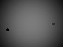
Fig.: Basal level ocular movement control training – fixation position and eye guidance in saccade, in horizontal direction, simultaneously training of advanced level of eye movement control – smooth pursuit eye movement during transition on a line / horizontal – vertical. Course of exercise: Animation of moving point from left to right with the transition to a line below. Points gradually appear on the left side, after finishing the motion to the other side they slowly disappear and at the same time a new point appears on the line below and the process repeats itself.
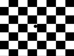
Fig.: Stimulation at the level of controlled transitions of ocular movements in the visual field in various directions – visual attention training in advanced perception range, during the observed point position change, during differentiation of figure and background. Course of exercise: Animation of point moving from left to right with unstable contrast of background. The point disappears and appears while moving on the chessboard. Patient observes and detects the point.
- Hearing
2.1 Neuroanatomy and physiology of hearing.
The ear is a very sensitive sensory organ which perceives sounds. Sound is a longitudal mechanical wave of the environment perceived by a person in the range of 16 – 20000 Hz. Sounds can be generally characterized by their frequency, pitch, timbre and duration. Sounds are harmonic and non-harmonic; every natural harmonic tone consists of a basic frequency and higher harmonic tones. Sound wave captured by the auricle passes through the ear canal and ends on the eardrum, which makes the lateral wall of the middle ear cavity. The auricle shape allows the directionality of hearing besides the capturing of the signal. Vibrating eardrum transmits the sound wave to the system of middle ear bonelets – malleus, incus and stapes. The last bonelet – stapes has its base located in an oval opening, which is part of the wall between the middle ear and the membrane labyrinth of the inner ear. The middle ear basic function is to transmit the acoustic signal from the large area of the eardrum to the small area of the oval opening, where the intensity of the signal is increased (Novák). Another mechanism of intensity increasing is the lever system of bonelets.
To transmit sounds efficiently through the middle ear, its condition is very important. Middle ear should be filled with air of the same pressure as the air in the surrounding environment. Ideal pressure conditions in the middle ear are facilitated by the Eustachian tube which enters the nasopharynx from the middle ear and allows balancing the middle ear pressure with the pressure of the environment. The middle ear also consist of these muscles – m. tensor tympani (innervations of n. trigeminus) attached to the handle of the malleus and m. stapedialis (innervations of n. facialis), which is attached to the stapes. These muscles change the solidity of the entire system of middle ear bonelets by changing their tension and thus the ear is reflectively protected against strong sounds – e.g. stapedial reflex (Hybášek).
The stapes with its base attached in the oval opening may be considered a piston, which vibrates the endolymph of the cochlear labyrinth membrane. The endolymph vibrates the tectorial membrane of Corti organ and thus excites the sensory cells. Corti organ is confine by Hensen cells and basilar membrane besides the tectorial membrane. The actual Corti organ consists of sensory epithelium, consisting of several types of cells. Cells important for hearing are inner and outer hair cells, which perceive the transmitted frequency vibration of the enolymph. (Note: Other types of cells are Dieters, (phalangeal), Claudius, Boettcher, inner and outer Corti pillar cells). Hair cells consequently convert the mechanical wave to an electrical impulse. The quantity of outer hair cells is in the ratio 3:1 to the inner hair cells, but the inner hair cells are in contact with the auditory path neurons. These neurons are myelinized and are more important to hearing. Outer hair cells are innervated by non-myelinized neurons (Husák, Kachlík). Corti organ has a tonotopic arrangement, i.e. every part of the cochlea allows perception of only a certain frequency. The sound wave decomposition to individual frequencies is caused by the actual shape of the cochlea and the hydrodynamic properties of the Corti organ. At the base of the cochlea there are perceived the highest frequencies and at its top there are perceived the lowest frequencies. Perception of sound intensity is set by the sound wave amplitude magnitude. The greated is the amplitude the greater is the area of hair cells reacting and the sound is perceived louder. Functionally the sound can be imagined as mechanical excitation of hear cells of certain areas of the cochlea, which is then converted to an electric impulse and transmitted using the auditory path to the brain stem and other higher stages of CNS.
Besides the described afferent neurons transmitting the signal from the ear to CNS, there are also efferent neurons, which bring fibres of the brain stem olivary complex to the inner ear (Husák, Kachlík). These are strongly myelinized fibres from medial olivary-cochlear bundle crossing the central line, which ends in the outer hair cells. These fibres inhibit or reduce the movement of outer hair cells and thus decrease sensitivity of Corti organ of the cochlea to sound. Non-myelinized mostly non-crossing fibres of the lateral olivary-cochlear bundle end in inner hair cells. It is assumed that their activities increase the level of necessary stimulus for the inner hair cells excitation. Functionally, the brain stem evaluates some sounds as disturbing and suppresses their perception. This tunes the ear to a certain signal as dominant and suppresses the surrounding noise/background; it is the so called selective hearing.
The actual auditory pathway has 4 neurons. The first neuron consists of bipolar cells, the body of which lies at the bottom of the inner ear canal in ganglion cochleare. Dendrites start in Corti organ at the base of hair cells, which react to the wave of endolymph. Axons compose thenervuscochlearis and they continue through the lemniscus lateralis and end at the bridge of the brain stem. Switching to the second neuron of the pathway in the nucleus cochlearis happens here. Axons of the second neuron compose the lemniscus lateralis and mostly cross to the other side and pass through the midbrain into coliculus inferior. There occurs switching to the third neuron. Its axons go to the corpora geniculata medialia of thalamus and switch to the fourth neuron, which axons compose radiatia acustica and pass into the primary auditory area of gyrus temporalis transversi (Heschl’s gyrus, area 41, 42) of the temple lobe. Subsequent analysis of the signal we hear is processed in the Wernicke area (Dubový, Jančálek).
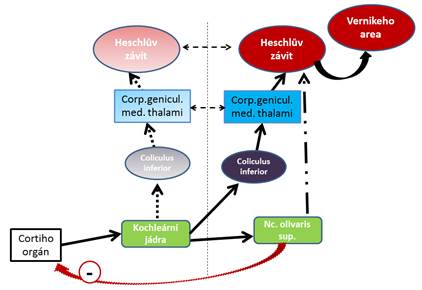
Fig.: Auditory pathway diagram, in deep colours there is cross-guided signal of speech character
2.2 Hearing in the context of central linguistic mechanism
Neural structures of Wernicke’s area are connected to the sensory regulation of speech/mechanism of understanding, but simultaneously it is assumed that they also compose the basis for inner linguistic concept formulation. This process can be understood that apart from the “pure understanding” the basic concept of answers which is primarily governed by Brock area is also created in Wernicke’s area. From the point of view of linguistic information transmissions, important association paths exist between frontal and temporal speech-linguistic areas (Lowe, Webb, 2009). Fibres connecting Wernicke’s area and Brock area (where the motoric programming of articulation is centred) are called fasciculus arcuatus. It is a long subcortical association tract, which connects rear and frontal linguistic areas of the brain; it transmits and circulates information from these areas back and forth. This process is the foundation of short-term auditory memory/phonological loop. Phonological loop works with phonological information. Auditory and verbal information is stored here into working memory and constant repetitions of this information keep them in memory before they are replaced by new information. Keeping information in the phonological loop is facilitated by sub-vocal articulation, i.e. inner speech. It is also called “articulation loop” (G.Hitch, A.Baddeley in Jošt). It is manifested as the inner voice, which resonates in our head, when we repeat the information we have heard, read for ourselves, repeat phone numbers we want to dial, etc. Phonological loop importance and its weakening are highlighted in the context of developmental disorders of speech, developmental learning disabilities or attention deficit disorder.
Terminology note: Phonem = basic/smallest auditory unit of speech. Distinctive features = minimum sound difference distinguishing semantic sounds (voiced: unvoiced … b:p). Phonological = transcription of audio signal of sound character into a semantic transcript of sound. From signal to meaning processing. Phonetic = audio signal of sound character generated by motoric realization using speech devices. From meaning to signal processing.(Palková)
2.3 Several important functions of auditory perception
2.3.1 Filtration
Alongside the second neuron interstitial cores are located: nucleus olivaris superior, corpus trapezoideum, and nuclei lemnisci lateralis, the most important auditory function of which is to filter acoustic noise and directionality of hearing. Filtration of acoustic noise occurs due to efferent nerves oriented into the ear, which are part of the entire auditory pathway and they influence our ability to transmit information from the ear to the cerebral cortex and they also influence the perception of sound at the inner ear level. To suppress useless noise signal olivary interstitial cores nuclei are used, out of which fibres lead to the inner hair cells of Corti organ of the inner ear and they are able to suppress their activity. Signal can be also suppressed alongside the pathway by decreasing the ability to switch
2.3.2 Location
To localize the sound, directionality of hearing occurs at several levels. Directionality of hearing is an ability the individuals learn during their development. For its perception, the auricle and its shape are important; and also the perception of sound signal through both ears, the more distant ear is delayed, unlike the signal coming to the closer ear. This delay is first evaluated in the brain stem, where the reflective connection to eye, head and body movements occur. At the same time direct signals of delay come from the olivary nuclei into the cerebral cortex, which takes preliminary steps to locate the sound accurately. The third factor which allows directionality is the acoustic pressure in both ears which is different. If one of the ears is disabled, the sound localization is possible with the help of the auricle and its shape to a certain degree.
Individual simple sounds are perceived even without the cerebral cortex involvement. The cerebral cortex is important for localization and recognition of sound time sequences. Time sequences are perceived in cochlear nuclei and in corpus geniculatum thalamu.
2.4 Ear and cerebral hemisphere dominance
Generally, the dominant hemisphere (the left hemisphere in most of our population) governs the interpretation of speech contents and the non-dominant hemisphere controls the holistic understanding of what we hear (recognizing emotional components, perception of music, etc.). To process signal adequately, the dominance of the relevant ear is necessary, the right ear for the left hemisphere and vice versa. If the dominance of the right and hemisphere is correct (right ear – left hemisphere, left ear – right hemisphere), the speech signal is processed in Heschl’s convolutions and Wernicke’s area of the dominant hemisphere, which controls the understanding of the contents (Koukolík). Emotional tone is mediated though the non-dominant other side. In the case of incorrect, i.e. ipsilateral dominance of the ear and hemisphere (left ear and left hemisphere, right ear and right hemisphere) the speech we hear is first interpreted “in a holistic” way through emotions, rhythm, melody, etc. and subsequently the contents is processed (Volemanová). If the ear or hemispehere dominance is non-existent, the situation is the least favourable and two signals coming from the opposite directions are delayed and there can be difficulties decoding the contents we hear which can even lead to defective speech generation (Lowe, Webb).
2.5 Principles and solutions with respect to auditory system stimulation and rehabilitation
Classical techniques of auditory perception development are mostly focused on working with words, their meanings and decompositions, i.e. at the lexical – semantic stress level and the level of sublexical stress, i.e. the phonetic – phonological differentiation level. (Terminological note: Linguistics uses the equivalent of “phonem consciousness”, i.e. the ability to recognize and to segment sublexical parts – phonems in the entire audio form of the word.) In practice, this development mostly occurs in the form of using various worksheets. On a smaller scale training of basic non-verbal sound recognition ability in the development of auditory perception is used, and if it is ever used, it is at the level of simple sound identification or development of auditory memory.
Authors of the article believe that principles of auditory perception rehabilitation stimulation are based on two basic needs. These are represented by the ability of sound perception and identification of our everyday surroundings and decoding of speech. From the point of view of acoustics these two lead to the necessity to distinguish the following modalities using sound: tonal/harmonic sounds, non-tonal/non-harmonic sounds, timbre of sound at identical and different pitches of the basic tone or noise, dynamics, sound duration, rhythm, melody, directionality, separating dominant signals from background. It is important to characterize the sound from the perspective of its generation and duration, in this category there are acoustically persistent and acoustically non-persistent sounds, these characteristics correspond with the fricative and non-fricative sound generation method in relation to speech. (Terminological note: In musical terminology, expressions non-percussive sound (persistent) and percussive sound (non-persistent are used). All the above mentioned modalities form the basis of speech, in its acoustic nature vowels are tonal phenomena, consonants are non-tonal/noise phenomena (voiced consonants have a tonal component). To recognize consonants and vowels it is necessary to consider an important acoustic characteristic, timbre, which corresponds with formant component. To distinguish consonants duration is also very important; there are persistent and non-persistent, explosively and fricatively generated sounds. Another important condition for quality processing of audio signal is the auditory memory (functional verbal-acoustic memory, also phonological loop or short-term working auditory memory). Auditory perception is a process of constant learning, when the individual creates verbal-acoustic links and due to them he assigns adequate meanings to heard sounds. Short-term verbal-acoustic memory disorders significantly worsen the development of speech as a linguistic system and the ability of motoric speech realization from the perspective of articulation and they make the learning process more difficult in general. (Note: Disorders of phonological consciousness and short-term verbal memory are the subjects of many research works: Mann, Liberman 1984; Liberman 1990; Bradley, Bryant 1985; Wolf, Bowers, Biddle, 2000; Waber at al., 2004. These research works focus mainly on the area of learning development disorders.
In the context of speech development and hearing impairments the authors have come to the conclusion that there should always be a basic training of auditory perception at a simple – acoustic level focusing on sounds which are necessary to differentiate and distinguish between sounds, words and environment sounds. In addition, it is suitable to include the development of directionality or selective hearing, i.e. the ability to distinguish between “figure and the background” of the sound we have heard. Such focused rehabilitation represents the basis for further efficient work at the phonetic-phonological level. Authors of the paper recommend and that the following sequence is used during rehabilitation/stimulation of auditory perception in clinical practice:
A/ Auditory perception development at non-verbal level/ with no verbal – acoustic links – in its simplified form it is work with three components. The first component is the training to distinguish and repeat generation/repetition of “simple sounds” using everyday objects which surround us or musical instruments of musical-therapeutic instrumentalist. In essence, it covers work with tone pitch, timbre, and methods of generation, duration, dynamics and rhythm. Furthermore, rehabilitation of modalities of directionality and spatial orientation are focused on. The last component is selective hearing, i.e. the recognition of “figure and background” of the sound we hear. (see below) These are the basics to work with before we move to the level of articulated speech and language/linguistic modalities. Only connected in this way it is possible/ advisable to develop the level of verbal-acoustic links. Important aspect is represented by the use of active sound or sound pattern repetition by the child. During the exercise other senses are gradually involved – sight, proprioception and touch. Simultaneously, this method works with auditory memory, which is important for further development of verbal-acoustic memory.
B/ Auditory perception development at verbal/lexically semantic stress level – when the auditory stimulation already works with words (words as semantic units). It is for example development of linguistic sensitivity working with rhymes and similarly voiced words, musical-motion work with words, rhythmisation of words, development of auditory attention at the level of hearing and reaction to given words, working with prosodic factors of speech – intonation, modulation including melody, speech pace (Term. note: From the point of view of phonetics and phonology it is suprasegmental level of speech), etc.
C/ Auditory perception development at sublexical/phonetic-phonological stress level – i.e. auditory stimulation at the level of word decomposition into sounds/phonems. It focuses on the development of phonematic hearing, i.e. auditory differentiation of distinctive features of sounds and the related “graphem-phonem correspondence” (i.e. ability to pronounce the recognized words as sound – give it sound while reading), development of auditory synthesis and analysis, etc.
2.5.1 Rehabilitation methods for auditory perception development on the non-verbal level
The rehabilitation methods described above are focused on the first area of non-verbal auditory perception development are designed for home exercises of parents and their children and in most cases do not require special aids. In the outpatient practice acoustic aids, musical instruments or PC programs can be used to enable sophisticated stimulation and rehabilitation. In auditory perception training if the hearing is impaired you can apply the same procedure at the beginning of rehabilitation. In the case of adults similar approach can be applied; however, it is advisable to adapt the method to their age, disposition and intellect. The following methods have been created and used by the authors of the paper..
I/ Non-persistent sounds drill: The child and the parent are sitting opposite each other with a small table in between them; there are three objects on the table. For tonality there are, for example, three various enamel or glass vessels (the difference lies in the tone pitch and timbre), to perceive non-persistent noise sounds small boxes from various materials can be used (differentiation of sounds is based on the perception of timbre). The parent uses a stick/ mallet to sound the individual objects and the child joins in. Then the child closes his or her eyes and the parent sounds one of the objects again, the child opens her or his eyes and attempts to “play” the same object. If the child is capable of recognizing all the sounds, you may proceed to repeating two in a sequence and subsequently three or four. In the given moment, apart from recognizing the individual sounds, auditory memory is stimulated. Another step in training is variety in the intensity of hits or addition of rhythmic pattern. the objects can be gradually changed so that their sound is more difficult to distinguish. It is important for the child to subsequently create actively the sound; since apart from perceiving the sound purely by ear they can also come to realize modalities through the hand movement, such as quiet – loud, fast – slow, and regular – irregular. Non-persistent non-harmonic sounds are the grounds for plosives perception. Tonal sounds are important to perceive vocals. The authors believe that using non-harmonic sounds are more important than using harmonic/ tonal sounds to stimulate sense of hearing with the aim to improve perception and to generate speech.
II/Persistent noise sounds drill: The procedure is similar to the previous practice; however, the source of sound is represented by crumpled paper, various types of polythene and cellophane. The parent rubs these objects or crumples them. When the sounds have been introduced to the child, the child closes his or her eyes and the sound is repeated, then the child opens the eyes and chooses the correct material from several options and repeats the sound. While practising, fast press in hand and long rustling can be distinguished. Persistent non-harmonic sounds form the basis for perception of e.g. sibilants.
It is important to stimulate/ rehabilitate distinguishing between non-tonal sounds of non-persistent and persistent nature to improve differentiation between consonants in the case of hearing impairment or auditory perception disorder in developmental dysphasia syndrome.
III/Directionality drill: This drill can vary and it is suitable for group rehabilitation. The basic scheme is the child with eyes closed sitting in the middle of a room. Another person slowly and quietly moves in the room while making one of the selected sounds (it is advisable to choose persistent sounds – rustle). The child points his or hand towards the sound direction while the other person is moving. In the case of error the child can check visually – opens his or her eyes and familiarize with the surroundings with the help of sight. Movement of more people with various sounds represents a harder level of the drill; in the case of small children it is advisable to use various colours to distinguish the objects (various colours of polythene, paper, etc.). The persons move quietly across the room and make sounds individually; the child is asked to determine its direction and match the person with the sound based on the colours). This drill can also be interpreted as the training leading to space realization, when the child is in the centre of space and hearing or visual checking works with grasp/ perception of space. Then, at the same time, drill aimed at rehabilitation of sensory deficiencies of integration is carried out.
IV/Directionality drill and dominant signal and background differentiation: The drill is carried out in the same way and variations like methods described in III to develop directionality. The only difference is that another secondary sound/ acoustic signal is added in the background. This sound is made by another person, or it is produced by the child when working with more “stress”. In the case the child makes the background sound, due to focus on the activity the background sound is dominant for the child and the main signal appears to be on the periphery. In this way significant rehabilitation stress is created. You can also agree on a word to be used as the dominant signal.
The above described methods can be introduced in rehabilitation plan in the case of serious speech impairments, hearing impairments or generally in the initial rehabilitation stages. Only after stimulation and mastering these nonverbal modalities, stimulation and development on the level of verbal stress is suitable in further rehabilitation stages. The sequence corresponds with the processes of understanding the world based on auditory perception in the course of development of the human being. At first, even speech is perceived by children nonverbally – acoustically. From the point of view of linguistics, in the early communication development of the child the first developmental stage of the adults’ speech “perception“ with further development proceeding to the stage of “understanding“ the adult’s speech. Before the 3rd month of age simple ability of the child torecognize and locate human voices and distinguish them from other sounds is created. Between the 4th and 8th months of age intonation, accent and segmentation ability is created – they recognize words in sentences they heard in isolation before. Between 10th and 12th month of age the child starts to understand words and is able to recognize various meanings. (Průcha, J., 2011).
Note: Auditory perception rehabilitation is dealt with at various places – Auditory Therapy Centres. The centres lay emphasis on auditory stimulation especially in the context of dyslexia and developmental disorders. These are e.g. Auditory Integrative Training in Westport, USA, The Listening Centre in Toronto, Canada, Dyslexia Research Laboratory of Dr. Kjeld Johansen, Denmark.